


Azure Biosystems
Designed for flexible choice in detection chemistry and samples, the Sapphire FL brings precise quantitation of nucleic acids and proteins.

The Azure Sapphire FL Biomolecular Imager delivers high-sensitivity, quantitative imaging for proteins, nucleic acids, and fluorescent samples. With versatile detection modes and intuitive software, it ensures fast, accurate, and reproducible results for Western blots, gels, and microplates. Compact, powerful, and reliable, it’s the ideal tool to accelerate molecular biology research.
Request for Demo Request for Quote
Flexibility with uncompromising performance
The Sapphire FL is the second generation Sapphire and ultimate biomolecular imager for FLEXIBILITY. With customizable and user-changeable laser and filter modules, it easily adapts to a lab’s changing needs and advancing research. The Sapphire FL offers customizable and user-changeable optical modules, 5–1000 μm resolution scans, a Z-plane range from -1.0 to +6 mm, five anesthesia ports for imaging living animals, chemiluminescence detection through a Chemiluminescence Module, and much more.
|
Unlimited Laser and Filter Wavelength combinations It features a unique, patent-pending design of interchangeable and customizable laser and filter modules, enabling a virtually infinite number of spectral combinations. A broad range of excitation and emission wavelengths, as well as phosphor imaging, are supported.
The Sapphire FL is the only biomolecular scanner in its class that reaches into the UV spectrum, allowing multiplexing of UV dyes with dyes in the NIR. Using the 375 Standard Optical Module, it is able to image common DNA stains, such as Dapi and Hoechst.
|
|
|
|
Application Flexibility The flexibility of the Sapphire FL guarantees you the freedom to confidently choose the best dyes and chemistry for any experiment. Custom optical modules can excite any dye at its peak, resulting in the highest possible signal for any fluorophore. In addition to fluorescence, the Sapphire FL also excels at imaging phosphor screens and chemiluminescence using the optional Chemiluminescence Module. |
|
Sensitive Fluorescent Detection High sensitivity allows femtogram detection of proteins labeled with common fluorescent dyes. No other system on the market matches the Sapphire FL’s coverage across the fluorescent spectrum. Extend your reach into the UV spectrum with the 375 Standard Optical Module. |
|
|
Plot showing linearity and linear dynamic range achieved when a carbon-14 standard was imaged using the Sapphire
|
Total Flexibility with Phosphor Imaging The Sapphire FL is a compact phosphor imager that incorporates exceptional visible fluorescent and near infra-red (NIR) scanning, along with true chemiluminescent and white light imaging, to make it a true lab workhorse. It is one of only two scanning systems in its class that offers phosphor imaging, a high sensitivity assay that requires radioactivity. The Sapphire FL is able to scan storage phosphor screens with exceptional dynamic range and image quality, thanks to the use of laser excitation and photon multiplier tube (PMT) detection. |
|
In Vivo Imaging The Sapphire FL imager makes imaging of live animals possible. Living mice or other small animals can be fluorescently imaged under anesthesia using any of the five built-in anesthesia ports. The wide imaging platform can accommodate specimens with depth (up to 4 cm), allowing small animals, plants, petri dishes, and other large biological samples to be imaged. |
|
|
|
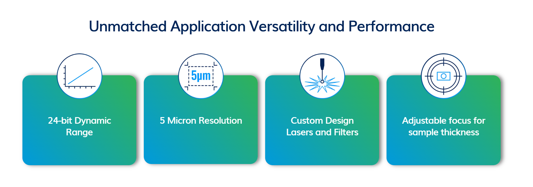
Distinguish between Subtle Differences in Expression with Extended Dynamic Range (EDR) Extended dynamic range allows you to image both bright and weak bands without experiencing saturation. This is ideal for the samples that feature strong and weak expressing proteins. EDR extends dynamic range to 24 bits of data. |
|
Adjustable Focus Some samples can be imaged on the glass (Western blots), while others are raised above the glass surface (96-well plates). The Sapphire FL can image them all. Some samples have multiple focal points within them, such as tissue slides and organs. The Sapphire FL can image those too. It offers a 7mm software-controlled adjustable focal plane, so you can scan your sample at multiple depths to ensure you see all your data has to offer. |
|
|
|
5-micron Resolution The Sapphire FL scans between 5 – 1000 µm resolution to ensure clear visualization of detailed samples. Image complex and minute samples, including tissue and microarray slides, with accuracy. Triage multiple slides at high resolution before microscopic analysis. Mouse lung tissue slide probed for vascular endothelial (VE)-cadherin (AzureSpectra 550 nm secondary antibodies) and smooth muscle actin (SMA) (AzureSpectra 650 nm secondary antibodies). Imaged on the Sapphire FL using the 532 and 638 Standard Optical Modules (red and green, respectively) at 5 µm. |
|
Chemiluminescent Imaging When You Need It An optional Chemiluminescence Module adds high-resolution, quantitative chemiluminescence and visible imaging. It allows you to express proteins with femtogram sensitivity and gives you the ability to capture color marker images. Western blot of serially diluted HeLa cell lysate in duplicate with marker, probed for Tubulin and Actin, detected with the Flash Western Chemiluminescence Kit. Visualized on the Sapphire FL Chemiluminescence Module. |
|
Visit https://azurebiosystems.com/products/sapphirefl/ for details.
Please sign in first.
Sign inCreate a free account to save loved items.
Sign in